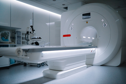
Multi Parametric Magnetic Resonance Imaging (mpMRI) – Prostate with PI-RADS scoring

What is multi-parametric magnetic resonance imaging (mpMRI) scan?
An mpMRI scan produces more detailed images of your prostate than a typical MRI scan. It does this by mixing different types of images. These scans provide information to your Uro oncologist regarding suspicious areas for cancer in your prostate.
Within the prostate, mpMRI measures water molecule mobility (called water diffusion) and blood flow (called perfusion imaging). This aids the Radiologist and the Uro oncologist in distinguishing between apparently diseased and healthy prostate tissue.
Why mpMRI scan is done?
An mpMRI scan is used to stage the prostate cancer. It tells if it is restricted to the prostate, or has spread to nearby organs like seminal vesicles, bladder neck, rectum or lateral pelvic walls. It also helps in staging the drainage as well as distant area lymph nodes in pelvis and retroperitoneum. It may give information of distant spread into bones or visceral organs like liver.
An MRI of the prostate can diagnose, evaluate and rule out conditions like:
- Prostatic abscess or infection (prostatitis).
- An enlarged prostate.
- Prostatic Carcinoma
How is Prostate Cancer diagnosed? | Medanta
Contrindications of mpMRI scan
- Claustrophobic patient
- Incompatible implants
What is Prostate Imaging Reporting and Data System (PI-RADS)?
The Prostate Imaging Reporting and Data System (PI-RADS) is used by radiologists to determine how probable a suspicious area is to be a clinically significant prostatic malignancy. The PI-RADS score ranges from 1 (no cancer) to 5 (cancer) (very suspicious). This scoring can be fused on software with MRI Fusion/TRUS Biopsy machines to perform accurate biopsies that pick up clinically significant prostate cancers.
Evolution to Biparametric MRI Prostate
What is Biparametric Magnetic Resonance Imaging (bpMRI)?
In patients with suspected prostate cancer, biparametric magnetic resonance imaging (bpMRI) of the prostate maybe done. This combines morphologic T2-weighted imaging (T2WI) and diffusion-weighted imaging (DWI). It is emerging as a viable alternative to multiparametric MRI (mpMRI) prostate for detecting, localising, and guiding prostatic targeted biopsy (PCa). It is less time consuming and helps resource optimisation in healthcare infrastructure.
Conclusion
For more information on prostate cancer related issues and best treatment options, visit Medanta Hospital and get medical assistance from our Uro Oncologist.


