Ductal Carcinoma in Situ (DCIS)

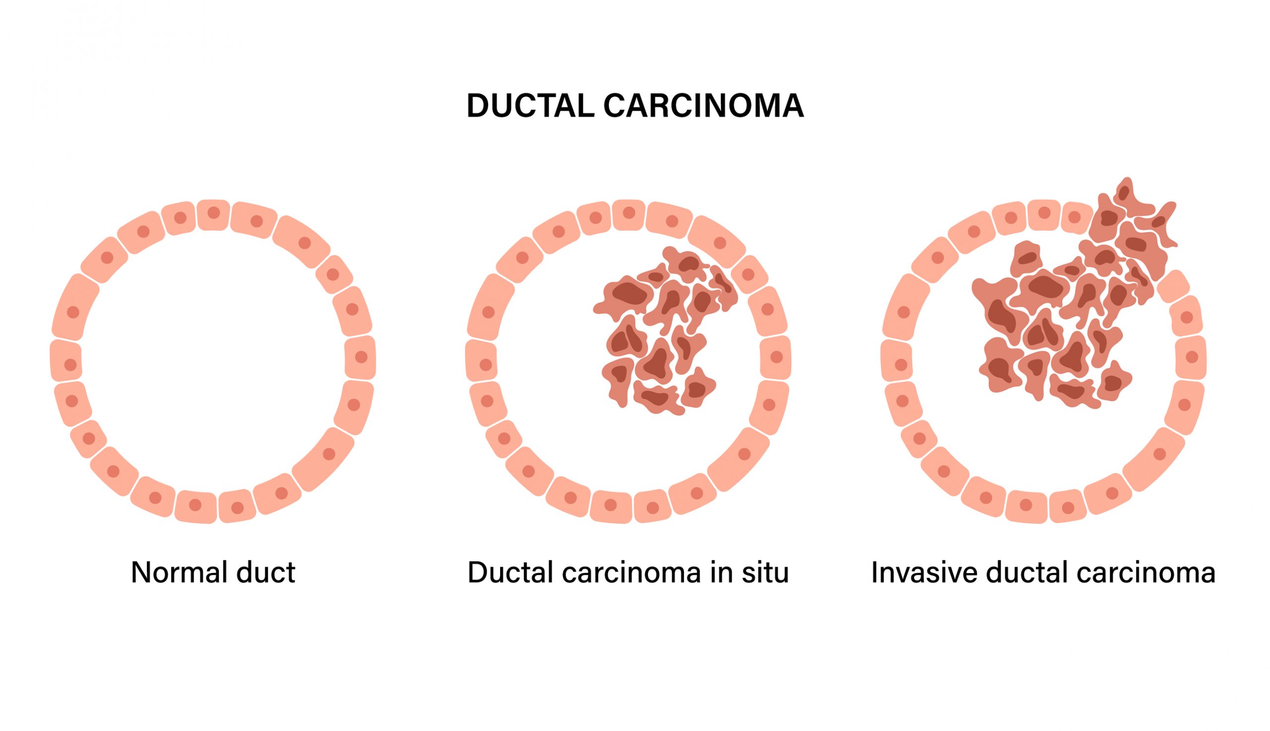
The presence of abnormal cells inside a milk duct in the breast is known as ductal carcinoma in situ (DCIS). The first type of breast cancer is known as DCIS. DCIS has a low probability of becoming invasive because it hasn't moved outside of the milk duct and is noninvasive.
To explore a breast lump or as part of breast cancer screening, DCIS is frequently discovered during mammography.
DCIS requires an evaluation and discussion of treatment alternatives even if it isn't an emergency. Surgery to remove whole breast or any other tumor tissue with radiation therapy is the treatment of choice.
Symptoms
There are no distinct signs of ductal carcinoma in situ, such as breast tenderness or a lump. The majority of instances are found through mammography before any symptoms appear. On occasion, a distortion of the breast tissue on the scan can be a sign of DCIS. DCIS most frequently manifests itself on a mammogram as new calcium deposits.
Itching or ulceration may appear once the cancerous cells begin to spread throughout the milk duct.
Men can get DCIS, and because they typically do not have routine screening mammography, the condition may manifest as a lump or a bloody nipple discharge.
A woman's or man's breast lump should be evaluated by a doctor soon because it may be aggressive cancer.
Stages
Since all DCIS is stage 0, your doctor will base their treatment recommendations on the DCIS grade. The grade indicates how closely the cells resemble normal breast tissue. DCIS is available in three grades:
Spend money to support free programs and resources for breast cancer survivors.
- Low-grade - DCIS cells are slow-growing and resemble healthy breast cells just slightly. The likelihood of low-grade DCIS returning (recurring) is lower than that of moderate- or high-grade DCIS
- Moderate-grade - Low-grade DCIS cells resemble healthy breast cells less than intermediate-grade DCIS cells, which also grow more quickly. Compared to low-grade DCIS, moderate-grade DCIS is more likely to recur, whereas high-grade DCIS is less likely to do so.
- High-grade - Compared to healthy breast cells, DCIS cells have a distinct appearance and develop faster. The likelihood of high-grade DCIS returning is higher than that of low- or moderate-grade DCIS. There are areas of dead cancer cells inside high-grade DCIS, which is referred to as comedo or comedo necrosis.
Diagnosis
A mammogram may detect anomalies in your breast tissue that a biopsy will allow your doctor to examine more closely.
Mammogram
In mammography, breast tissue is imaged using a low-dose X-ray. The small calcium deposits left behind by aged cells as they accumulate inside your milk ducts are known as breast calcifications. On mammography, calcifications show up as a shadow or white area. DCIS or other types of breast cancer may show aberrant cell proliferation, which is indicated by abnormal calcifications.
If a screening mammogram reveals any suspicious areas, your healthcare provider may decide to order a diagnostic mammogram in addition to the screening mammogram. With a diagnostic mammogram, you can see your breast tissue in greater detail with a diagnostic mammogram. It takes more time than a screening mammogram.
The following mammograms are used to find DCIS:
- 2D Mammograms - A traditional mammogram produces a two-dimensional (2D) image of your breasts by taking at least two images of them from various angles. The most common imaging method for identifying DCIS is a 2D mammogram.
- 3D Mammograms (Breast Tomosynthesis) - A three-dimensional (3D) mammogram creates a view of your breast from multiple images. Particularly in dense breast tissue, this type of mammogram detects breast cancer more reliably than conventional mammograms.
Biopsy
To verify that you have cancer cells in your breast, your doctor may do a core needle biopsy. To obtain a sample of the aberrant breast tissue, your doctor will put a giant needle into your breast during this surgery. A pathologist examines the lab-grown cells for any indications of malignancy.
Your doctor can use imaging to pinpoint the exact location of the abnormal tissue. During your biopsy, they might utilize an X-ray or an ultrasound. Breast biopsies that are guided by ultrasound are referred to as "ultrasound-guided biopsies." Stereotactic breast biopsy refers to a biopsy that makes use of X-rays, much like a mammogram.
Treatment:
Despite the fact that DCIS is not an aggressive or rapidly spreading cancer, it is crucial to get treatment. Without therapy, some types of DCIS may progress to invasiveness. This indicates that cancer has spread into the breast tissue around your milk ducts.
Breast-conserving surgery (lumpectomy) combined with radiation therapy or a mastectomy are the two most popular therapies for DCIS.
Conclusion:
The presence of abnormal cells inside a milk duct in the breast is known as ductal carcinoma in situ (DCIS). DCIS is the earliest stage of breast cancer. DCIS is noninvasive, which means it hasn't spread outside of the milk duct and is unlikely to become invasive.


