Bronchiectasis

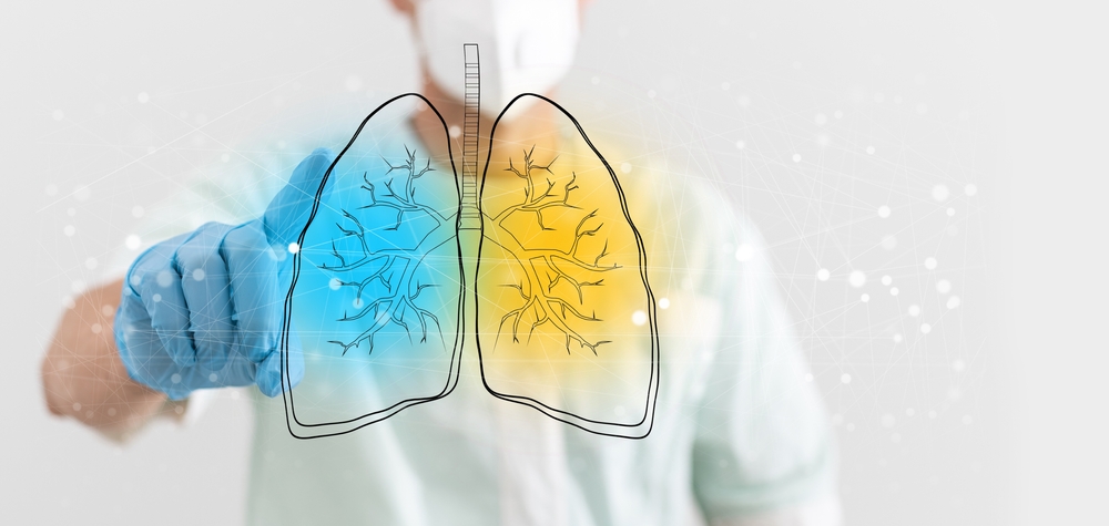
When you have bronchiectasis, your lungs' bronchial tubes are permanently enlarged, thickened, and damaged. Because of these damaged airways, mucus and germs can gather in your lungs. As a result, the airways often get infected and blocked.
Bronchitis cannot be cured, although it is treatable. With therapy, you can often lead a normal life. However, to preserve the oxygen supply to the rest of your body and stop additional lung damage, flare-ups must be treated right away.
The mucus layer and small protrusions (cilia) on the cells that line the airways serve as defenses. The thin liquid coating of mucus ordinarily covering the airways is moved by the back-and-forth motion of these cilia. This mucus layer's harmful contaminants and germs are transported to the throat, where they are coughed up or ingested.
Areas of the bronchial wall are harmed and develop chronic inflammation, whether the airway damage is direct or indirect. The afflicted airways enlarge (dilate) and generate tiny sac-like outpouchings that resemble tiny balloons as the inflammatory bronchial wall loses its elasticity. Additionally, inflammation causes increased secretions (mucous).
Causes:
Inflammation or recurrent infections of the airways are common causes of bronchiectasis. It begins in childhood, usually followed by a serious lung infection or the inhalation of some foreign item.
The most commonly observed causes of bronchiectasis are as follows:
- The build-up of thick, sticky mucus in the lungs of patients who have cystic fibrosis
- Autoimmune diseases including Sjogren syndrome and rheumatoid arthritis
- Allergic lungs problems
- Malignancies connected to leukaemia
- Immune system disorders
- First-degree ciliary dyskinesia
- non-tuberculous mycobacterial infection
In addition, the infection and inflammation may penetrate the lungs' tiny air sacs called alveoli, where they may result in pneumonia, scarring, and the degeneration of lung tissue. The right side of the heart may eventually experience strain as a result of trying to pump blood through the changed lung tissue in cases of severe scarring and lung tissue loss. A kind of heart failure termed “Cor Pupulmonaleresults from right-sided cardiac damage.
Chronic respiratory failure is a term used to describe very serious (advanced) cases of bronchiectasis, which are more common in low- and middle-income countries and in people with advanced cystic fibrosis. These cases of bronchiectasis can significantly impair breathing and lead to unnaturally low amounts of oxygen and/or elevated amounts of carbon dioxide in the blood.
Bronchitis and bronchiectasis:
Similar symptoms have been observed in both bronchitis and bronchiectasis which include coughing up mucous. However, bronchitis is a transient illness that doesn't permanently expand your airways, but bronchiectasis does.
Symptoms:
The signs and symptoms of bronchiectasis include:
- Coughing out a lot of pus and mucous
- Recurring colds
- Stench-filled mucous
- Breathing difficulty (dyspnoea)
- Wheezing
- Coughing up blood (haemoptysis)
- Enlarged fingers and curled nails (nail clubbing)
There may be periods of time when your symptoms aren't as bad, followed by an exacerbation in which they worsen. However, symptoms of an exacerbation include:
- Extreme weariness (fatigue)
- Chills and fever
- increased breathlessness
- Sweats during the night
Diagnosis:
The most popular test for bronchiectasis diagnosis is a chest computed tomography (CT) scan because a chest X-ray does not give enough information.
This painless examination produces exact images of the structures in your chest, including your airways. The degree and position of the lung injury may be seen on a chest CT scan.
Moreover, your doctor will attempt to determine the origin of the bronchiectasis based on your history and the results of the physical examination after the chest CT scan has verified the condition.
Finding the precise reason can help the doctor address the underlying condition and stop the bronchiectasis from getting worse. Bronchiectasis can be brought on by a wide range of factors.
Overall, pulmonary function tests, laboratory testing, and microbiologic testing make up the majority of the examination for the underlying cause.
Your initial assessment will probably consist of:
- Sputum culture to look for bacteria, mycobacteria, and fungus
- A full blood count with differential examinations
- Immunoglobulin levels (IgG, IgM, and IgA)
Your doctor will request a sweat chloride test or genetic test if they have a suspicion of Cystic Fibrosis (CF).
Treatment:
Although bronchiectasis cannot be completely cured, the symptoms can be managed. Healthcare professionals manage infections and clean mucus to treat bronchiectasis. Your doctor may recommend medication or physical treatment, depending on the seriousness of your issue. You can also make use of mucus-removing medical equipment.
Treating the underlying illness may relieve your symptoms if bronchiectasis is triggered by it. Surgery may be advised by your doctor if you have a tiny region of bronchiectasis, however, this is uncommon.
Conclusion:
A condition in which the airways of the lungs become damaged, making it difficult to clear mucus.
Bronchiectasis can be caused by an infection or a medical condition like pneumonia or cystic fibrosis. Mucus accumulates and breeds bacteria, resulting in frequent infections.
Symptoms include a daily cough that lasts months or years and the production of large amounts of phlegm on a daily basis.
Physiotherapy and medication, such as antibiotics and drugs to loosen mucus, may be used in treatment.


